A common benthic genus. Some species make okadaic acid in the wild.
I find benthic Prorocentrum in sand samples from nearly every tank I look at. Most of the time they sit at low levels and most people never notice them, but they can bloom. I do not have enough data to say how hard a Prorocentrum bloom is to deal with, and aquarists report different experiences.
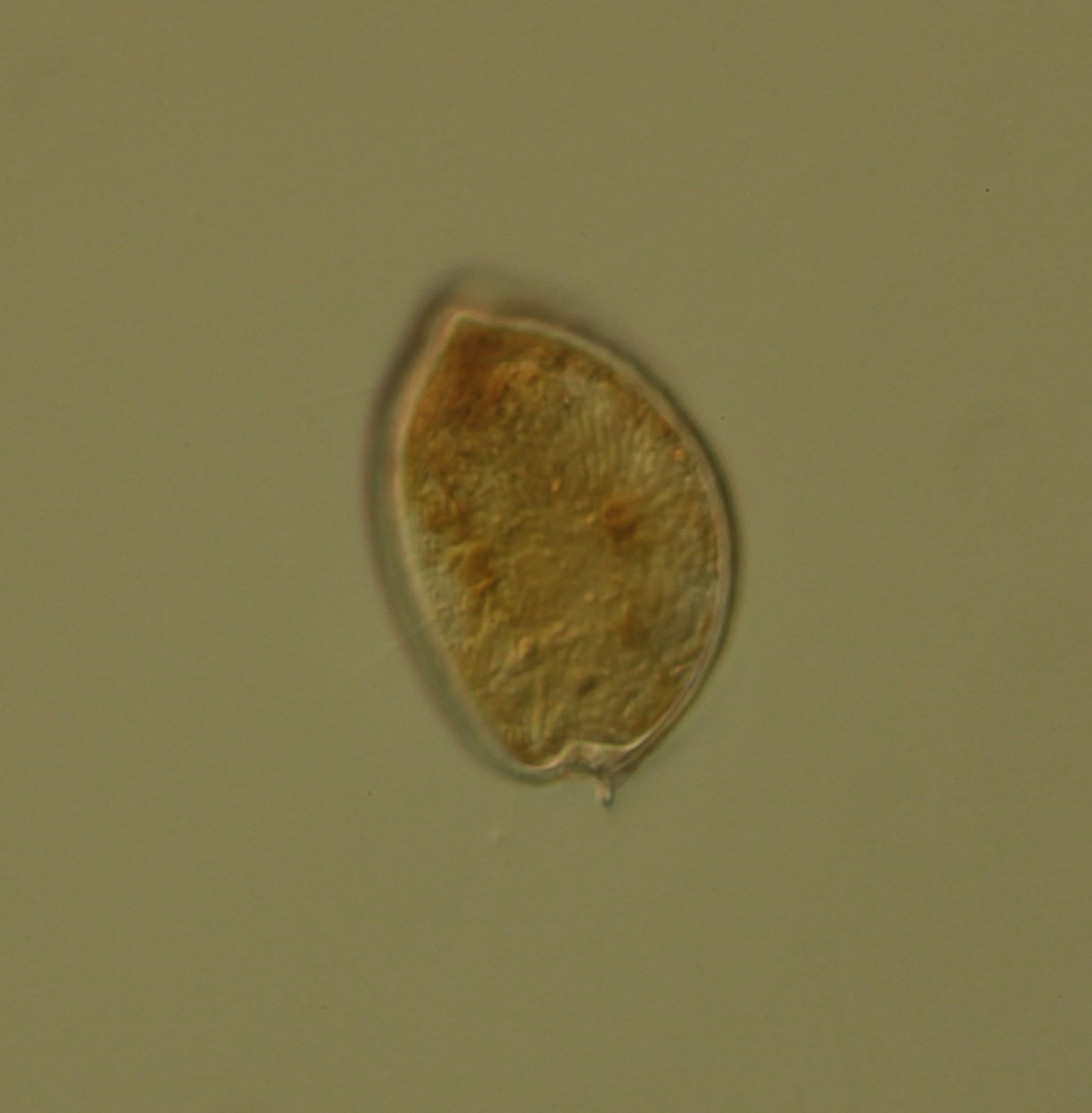
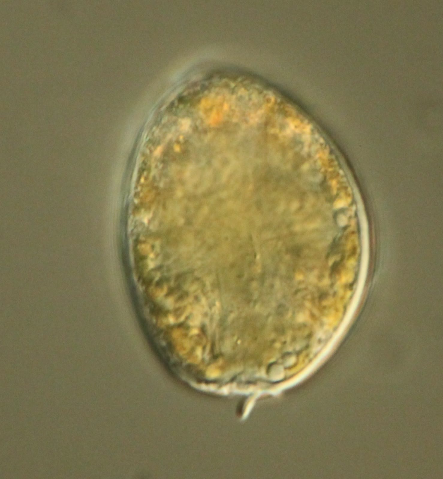
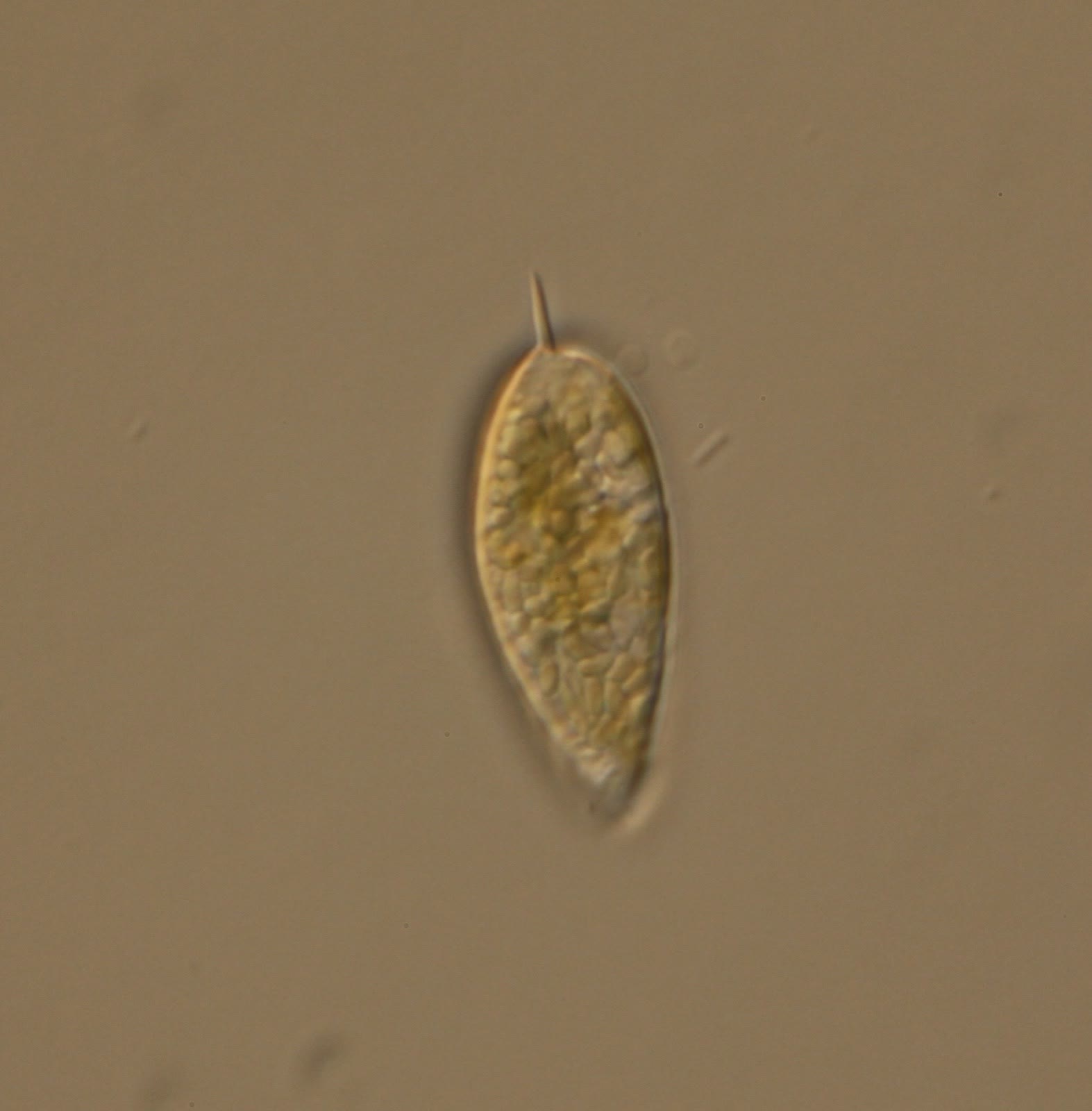
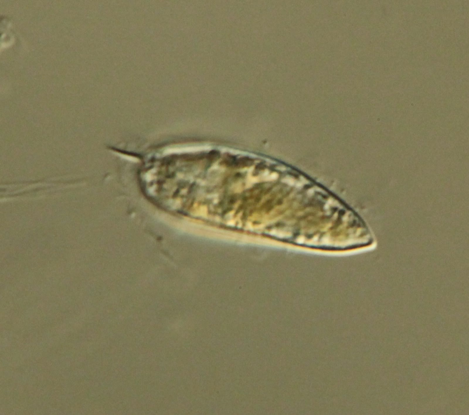
About toxins. Some Prorocentrum make okadaic acid, which causes diarrhetic shellfish poisoning in the wild. The genus falls into two major groups: a more symmetrical one, and one that is less symmetrical and often has a little spike (like some of the cells in the pictures below). The Prorocentrum we see in home aquaria are not thought to make okadaic acid, but it is very hard to say conclusively that any species does not make toxins, because it can be hard to get them to express toxins in the lab. I have not met anyone who has observed toxic effects from Prorocentrum in their aquarium.
How to recognize it
Size and shape. Individual oval, round, or heart-shaped cells, larger than small-cell Amphidinium (roughly 20 to 60 µm). Large-cell Amphidinium can be close in size, so use the other clues too.
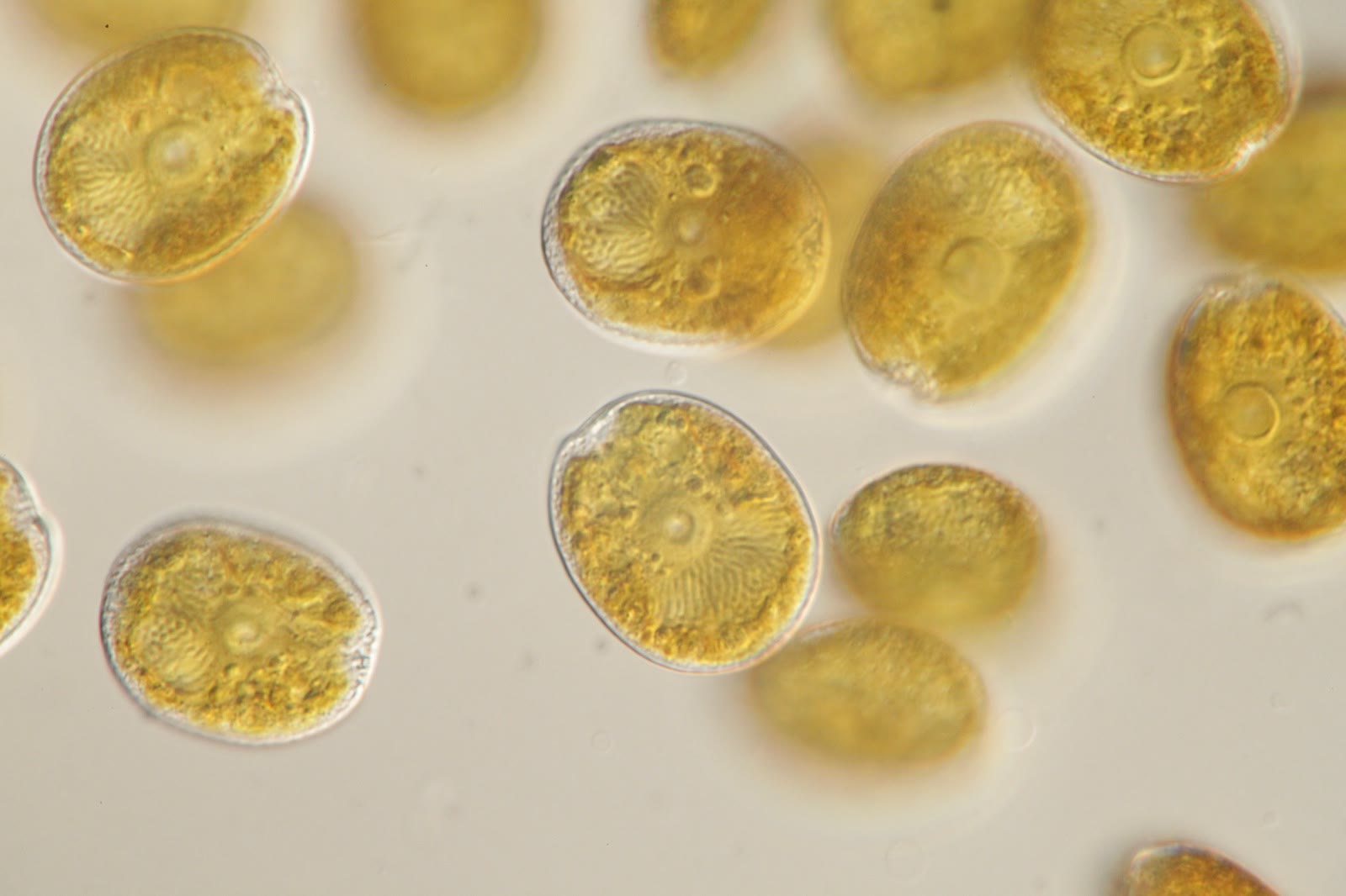
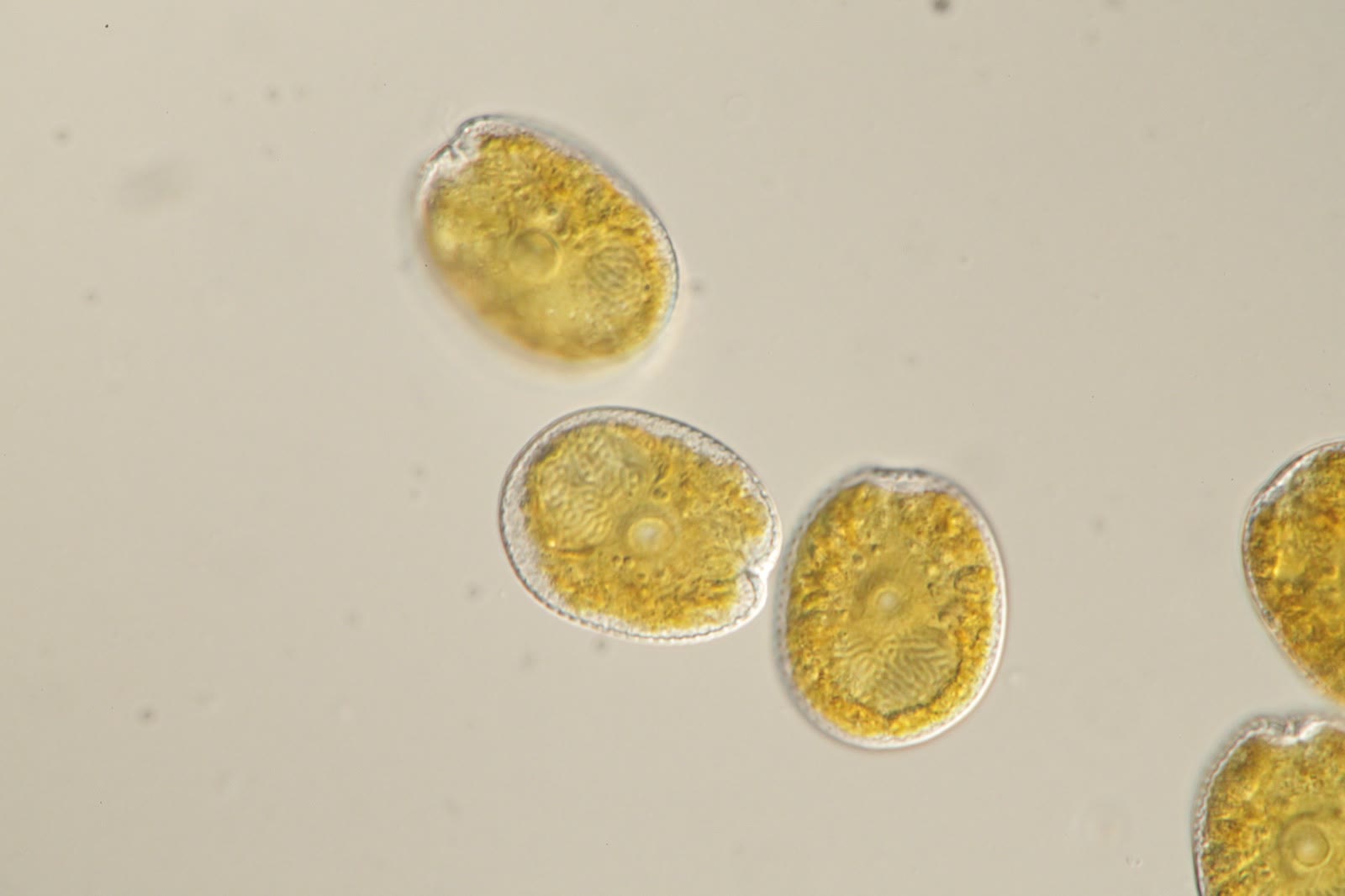
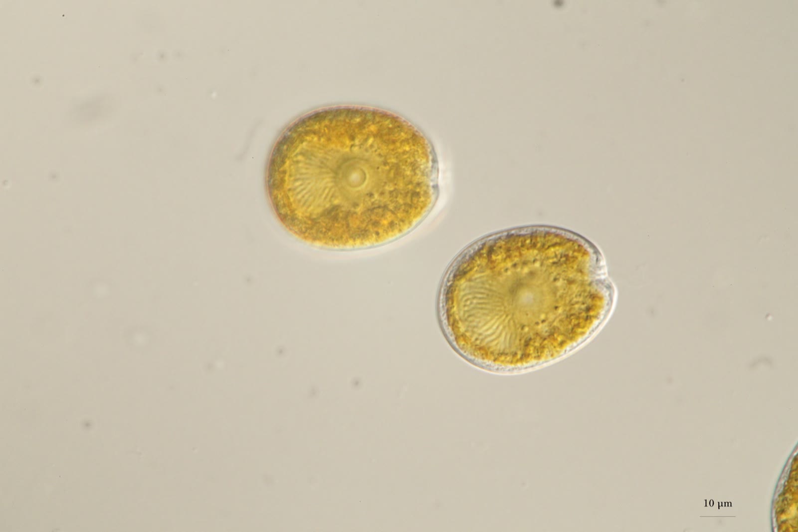
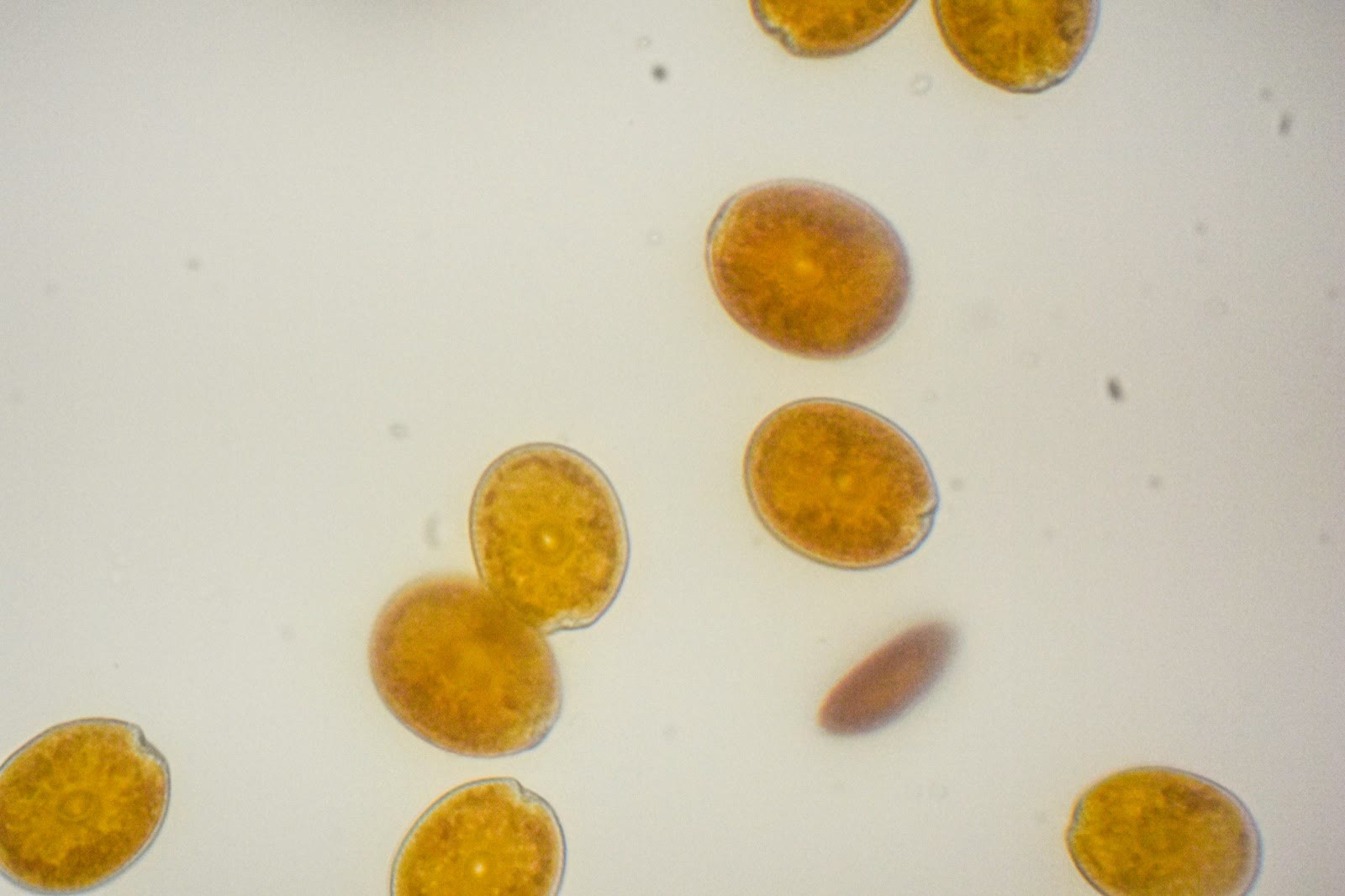
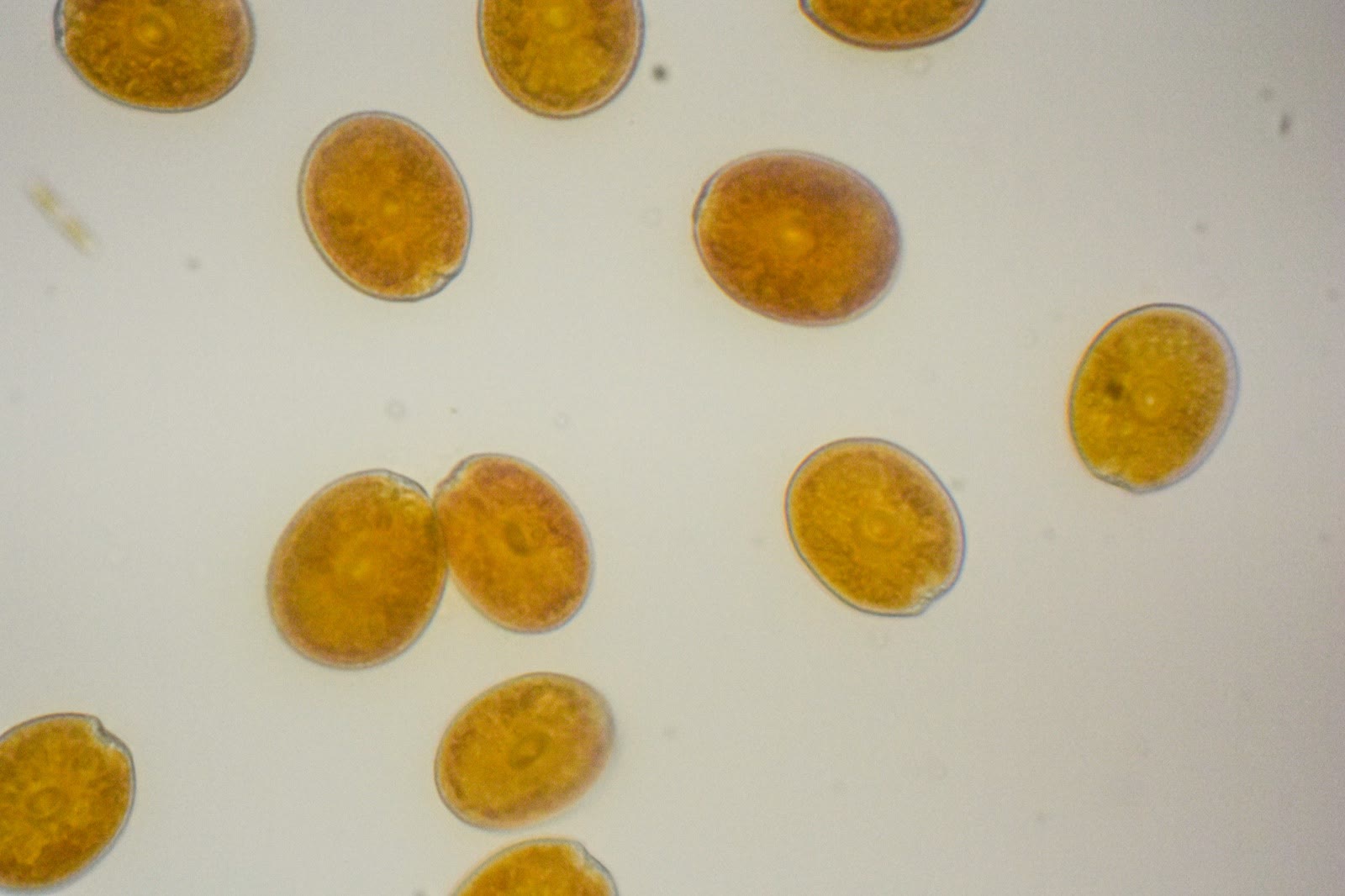
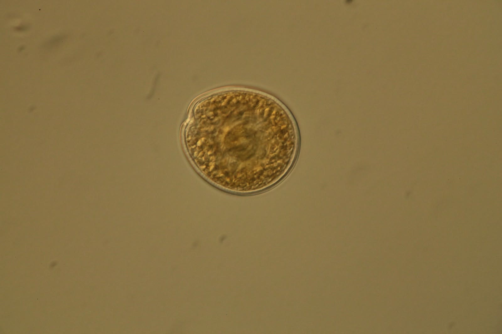
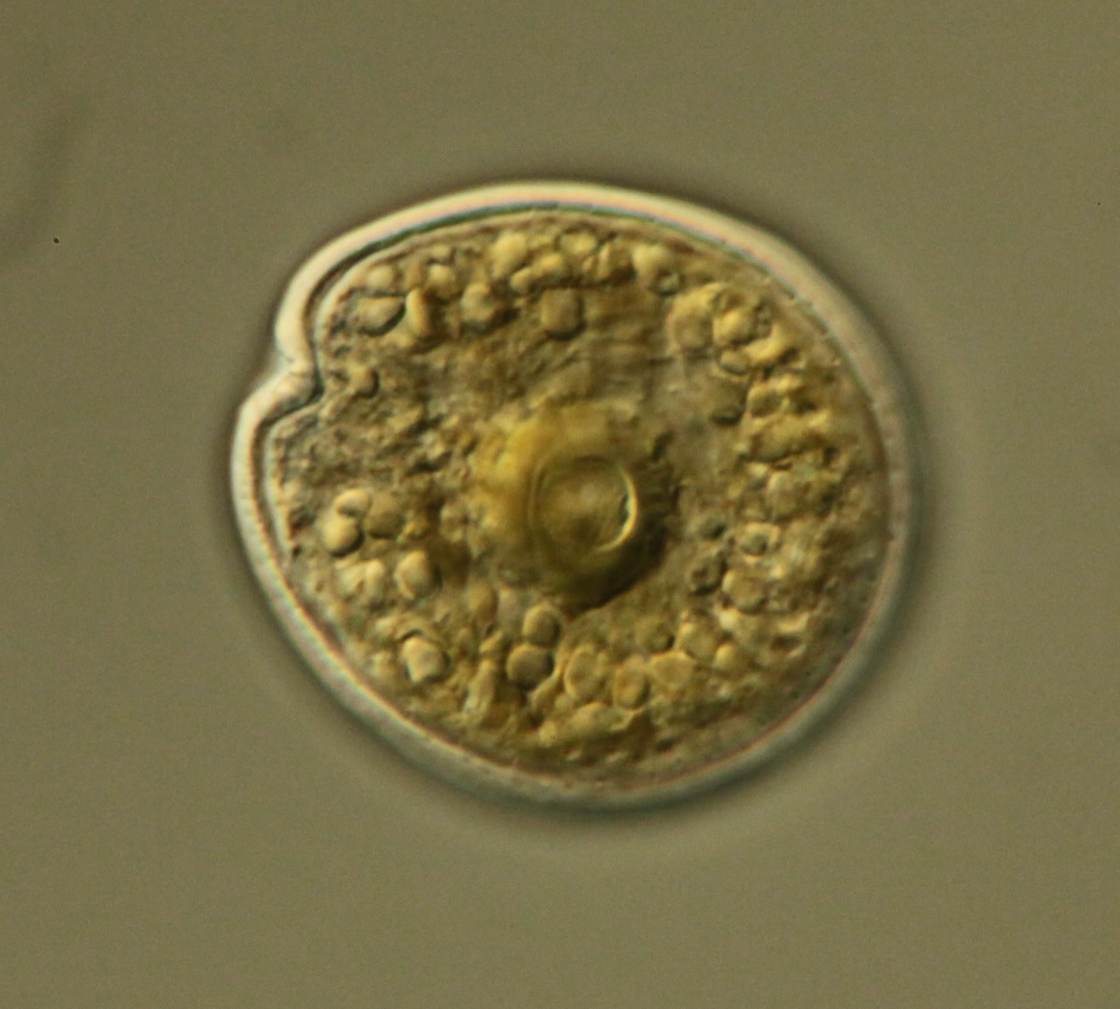
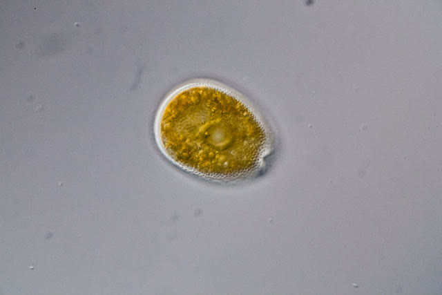
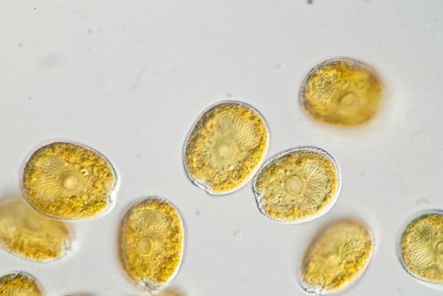
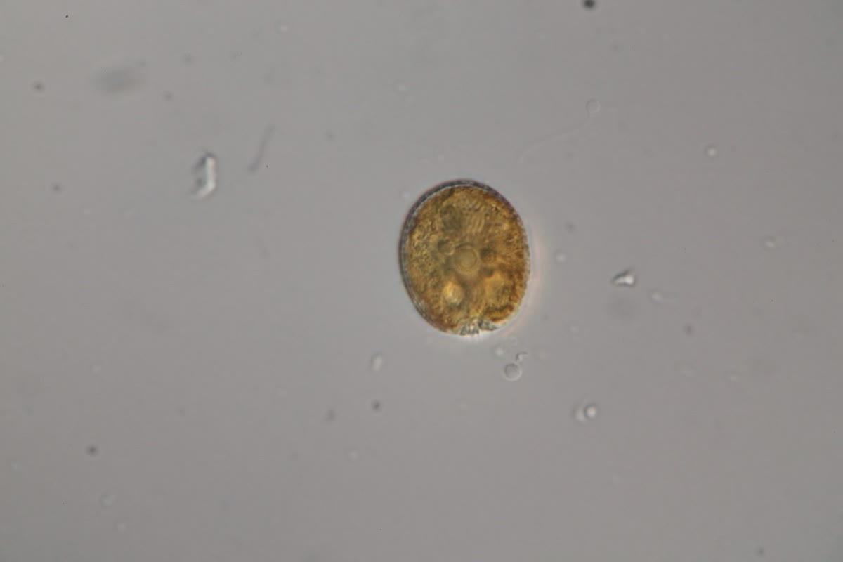
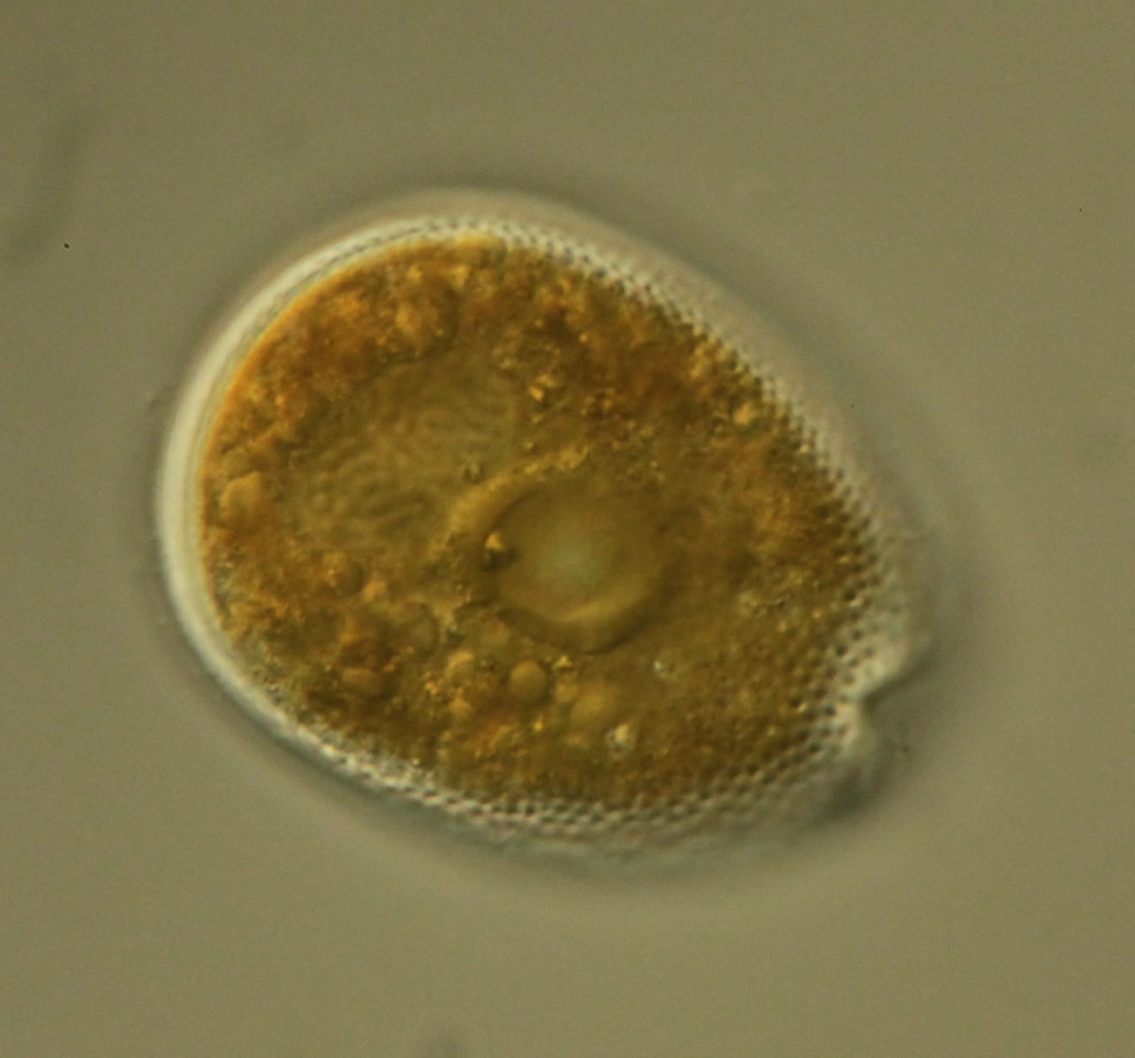
A large central pyrenoid. A big, perfectly round circle in the middle of the cell.
The nucleus (dinokaryon). It fans out behind the pyrenoid.
Movement. Usually none, or the tumbling swim typical of most dinoflagellates. They do not glide along surfaces like Amphidinium.
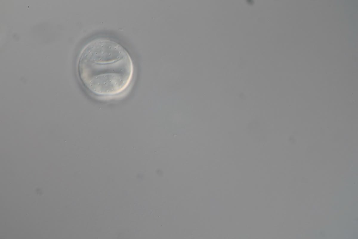
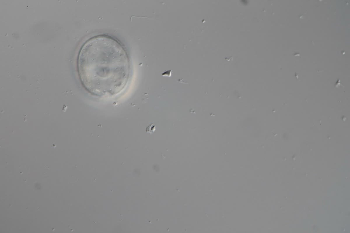
The apex.Prorocentrum cells have two halves (valves). One valve has a notch at the apex, and the two different flagella come out of pores in that notch.
People often confuse Amphidinium and Prorocentrum, since both are oval. Size, the pyrenoid, the motion, and the two narrow folds at the top of an Amphidinium cell are how to tell them apart.
Prorocentrum: mostly two big plates (valves). The grooves have shrunk into a notch at the apex, and there is no upper half.
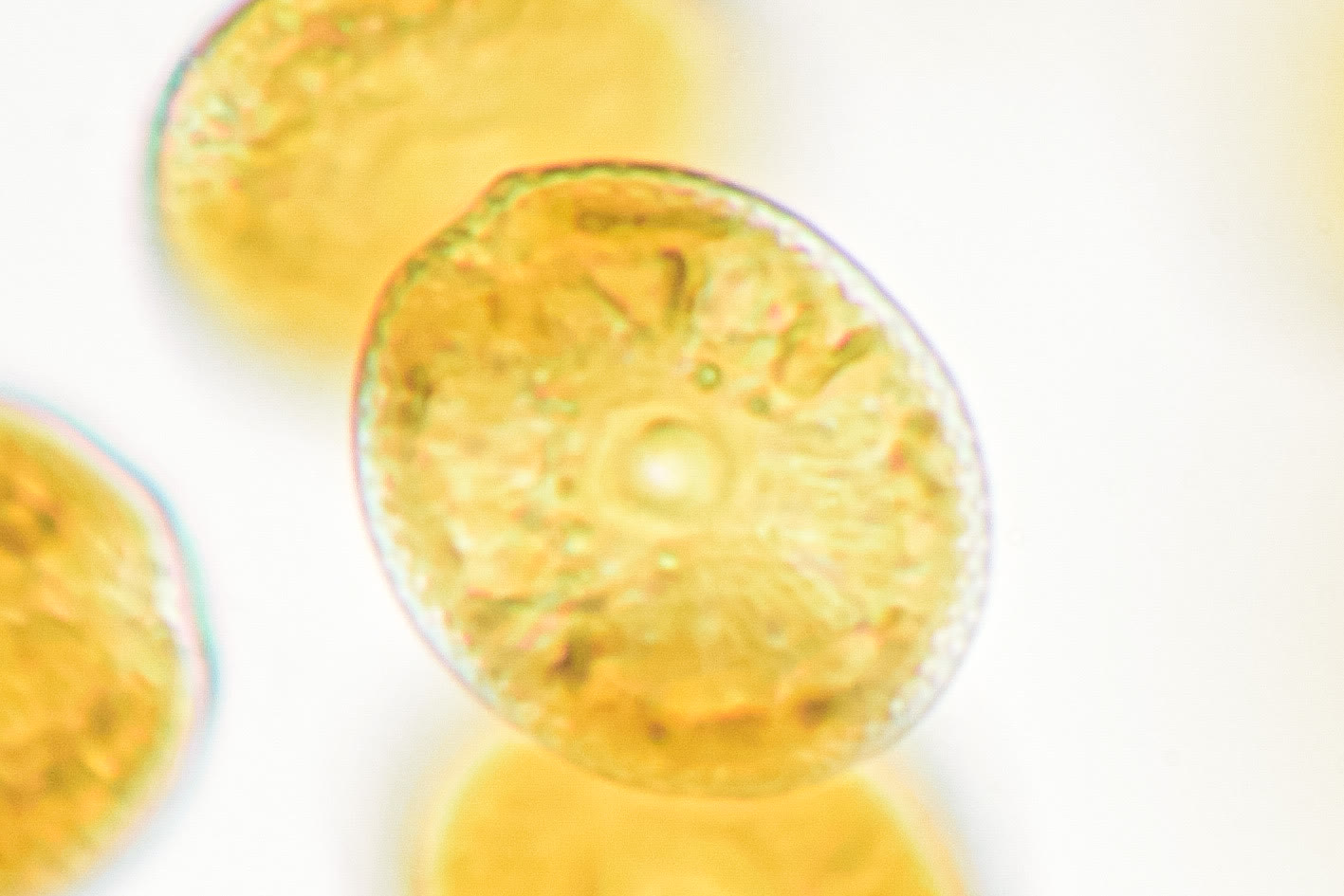
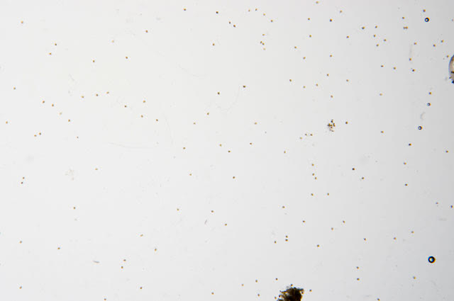
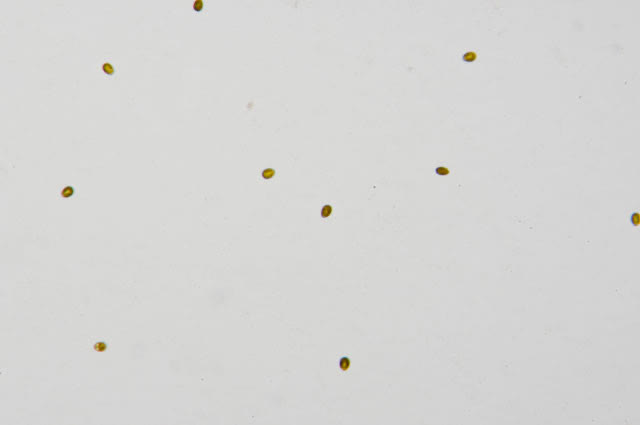
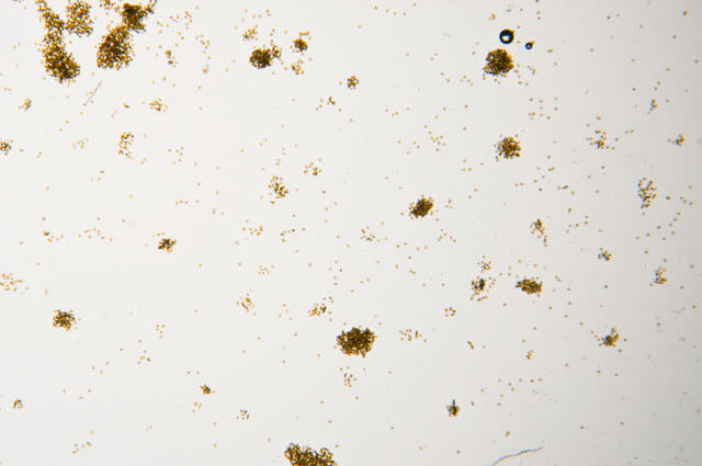
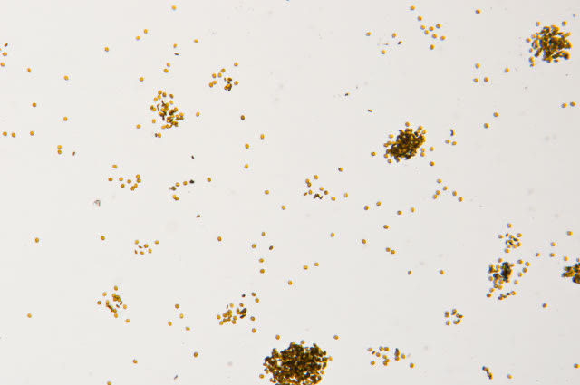
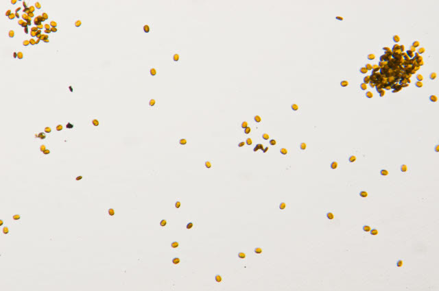
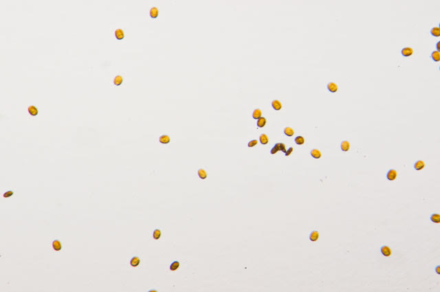
An undescribed species of Prorocentrum isolated from a marine aquarium, 630X. A large central pyrenoid is visible and the dinokaryon fans out below it.Same isolate, 630X. The pyrenoid is the large round structure in the middle of each cell.An undescribed Prorocentrum from a home aquarium (collected April 2012 by a marine aquarium hobbyist), 630X on a Zeiss Axioskop with a Canon EOS 5D Mark II. The cell rests on its left valve while the microscope is focused through the right valve, which is missing a notch at the apex. In Prorocentrum taxonomy the plates arranged in that notch matter, and the two different flagella emerge from pores there. The upper cell has the pyrenoid in focus; the lower cell has the dinokaryon in focus. The suture between the valves shows as a halo around the lower cell.Prorocentrum in close-up: oval cell with a round central pyrenoid. Image courtesy of a hobbyist.A field of Prorocentrum cells. Image courtesy of a hobbyist.Prorocentrum cells, a second field. Image courtesy of a hobbyist.
More species
Prorocentrum levis
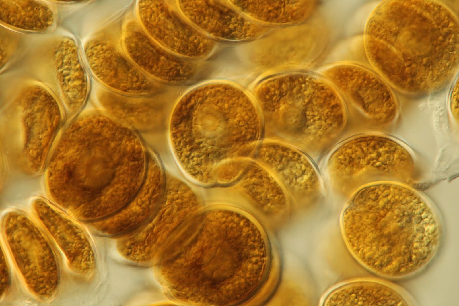
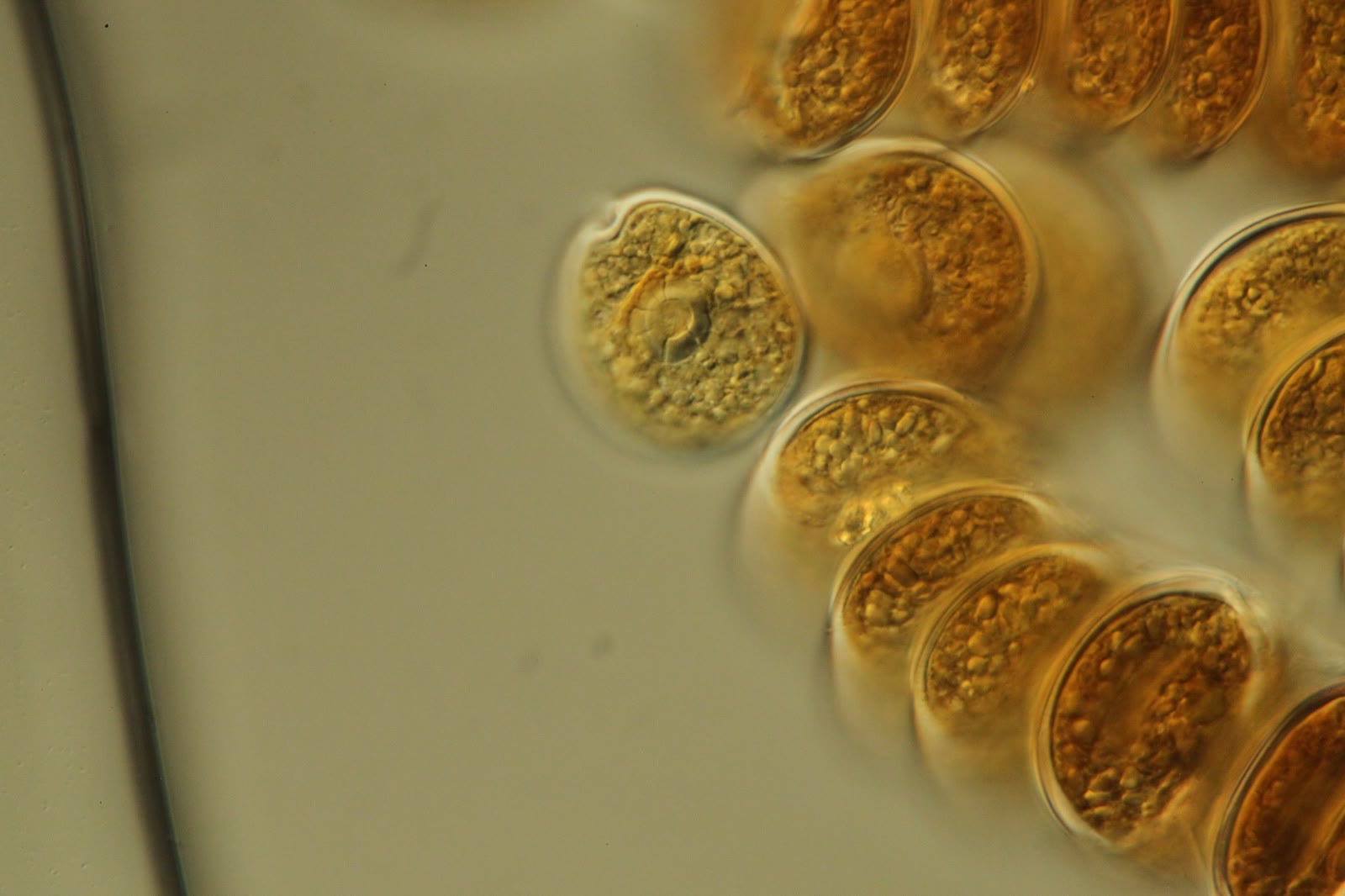
Often seen in stacked clusters, with large central pyrenoids that look segmented.
Stacked clusters of Prorocentrum levis. Large central pyrenoids are visible.Prorocentrum levis at 630X. The central pyrenoid is segmented.Prorocentrum levis at 630X.Prorocentrum levis at 630X, cropped.
Prorocentrum hoffmanianum
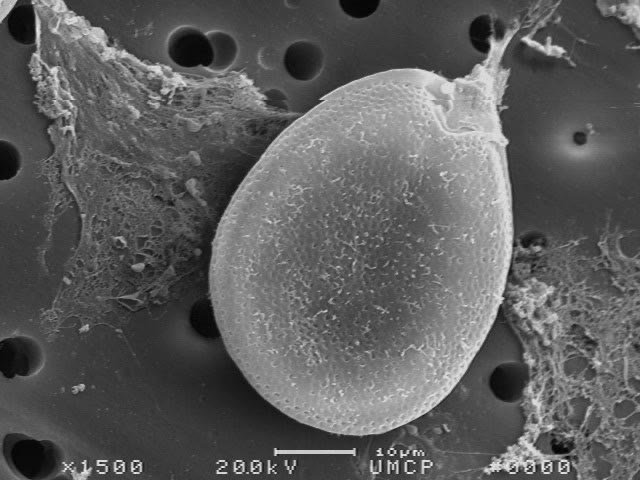
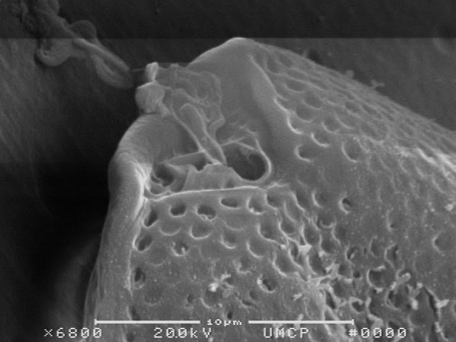
Light micrographs from low to high magnification, then scanning electron micrographs. Species identification of Prorocentrum often depends on the pores in the plates and the plates in the apical notch, which you cannot see with a light microscope.
Prorocentrum hoffmanianum at 10X magnification.Prorocentrum hoffmanianum at 40X magnification.Prorocentrum hoffmanianum at 630X magnification.Prorocentrum hoffmanianum, scanning electron micrograph at 1500X. Scale bar 10 µm.Scanning electron micrograph at 6800X showing the apical notch and the pores in the thecal plates of P. hoffmanianum. Scale bar 10 µm.
An undescribed Prorocentrum from an aquarium
This is the isolate shown above, collected from a hobbyist's aquarium and photographed at several magnifications.
Prorocentrum sp. at 10X magnification.Prorocentrum sp. at 20X magnification.Prorocentrum sp. at 40X magnification.Prorocentrum sp. at 60X magnification.Prorocentrum sp. at 630X magnification.
A newer aquarium: Prorocentrum arenarium?
In 2012 a hobbyist sent me this one, from an aquarium only a few months old. Snails and a sand-sifting goby would not graze on it, and he thought some snails died after grazing on it. I tentatively identified it as P. arenarium, but I could not be sure from pictures alone.
A Prorocentrum isolated from a new aquarium in 2012 (tentatively P. arenarium), living cell.Left valve of the same isolate.Right valve of the same isolate.
A variety from culture collections
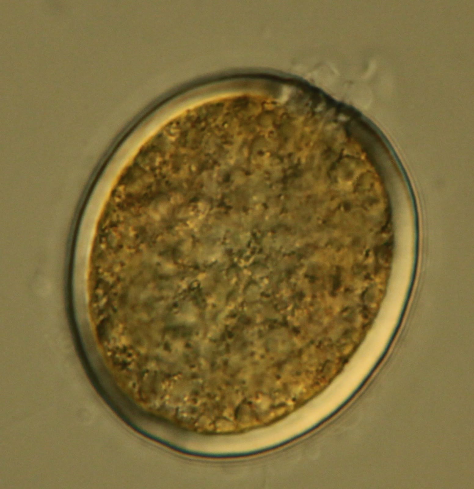
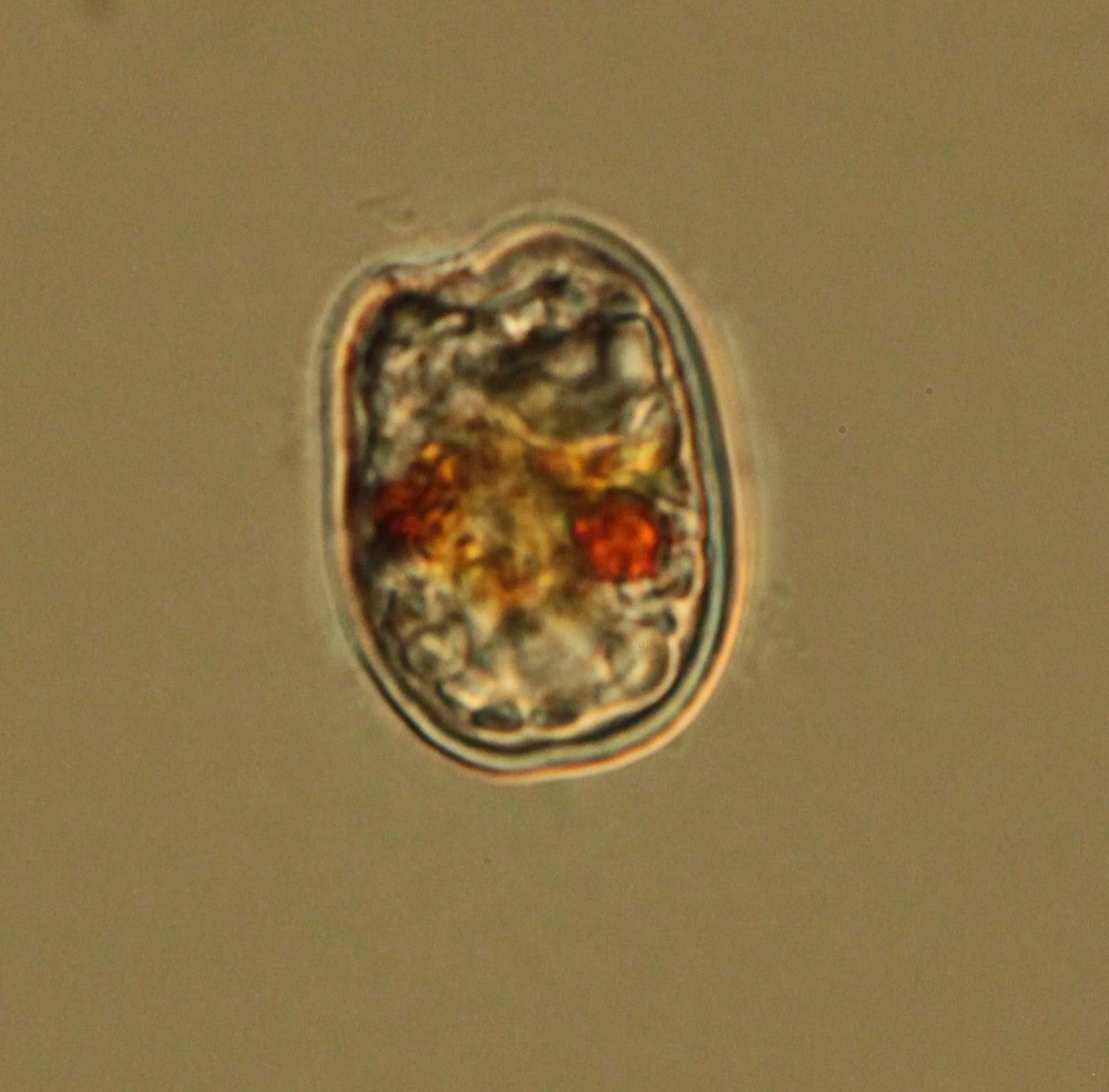
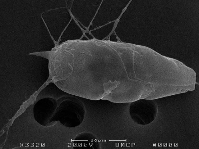
In 2011 I bought a variety of dinoflagellates, mostly Prorocentrum species, from culture collections. They show how different Prorocentrum cells can look, and that they are flat: some are pointed, some round, and some carry a spine. The species were not recorded in my notes.
A pointed oval cell, one of several Prorocentrum-type dinos I bought from culture collections in 2011. The species was not recorded here.Another culture-collection cell: a larger oval with a central pyrenoid and a row of pores along the edge of the plate.A paler oval cell. The notch at the apex (at the bottom of this picture) is where the grooves and flagella are crammed in.A slender, pointed cell with a spine at one end.A slender cell with a spine and a trailing flagellum.A round cell with a thick wall. Cultures often contain cells in different stages like this.A cell with orange-red bodies inside, as seen in older or stressed cultures.
Scanning electron microscope views
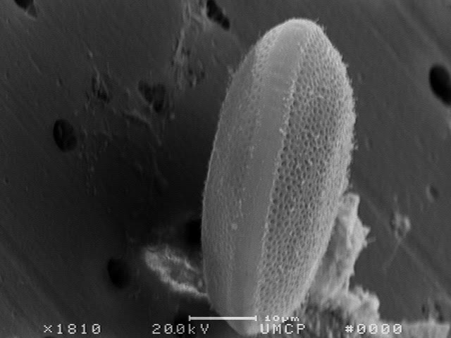
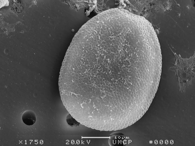
The scanning electron microscope shows what you cannot see in an ordinary microscope: the pores in the plates, and that the cell is a flat disc when seen from the edge.
A scanning electron micrograph of a Prorocentrum seen from the edge. It is flat, a thin disc made of two plates. Scale bar 10 µm.The same kind of cell seen face on, with the pores in its plate. Scale bar 10 µm.A scanning electron micrograph of a pointed Prorocentrum with a spine at one end. Scale bar 10 µm.
Fluorescence
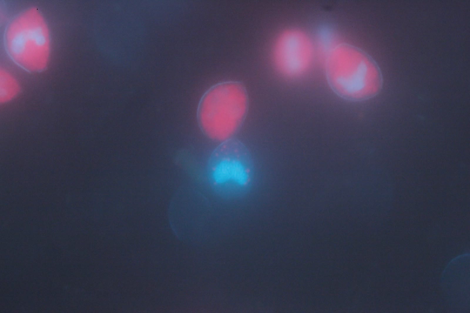
Epifluorescence microscopy of Prorocentrum micans stained with DAPI. Nuclei appear blue from the DAPI; chloroplasts glow red from autofluorescence. The characteristic bi-lobed nucleus is visible.
Video from another aquarist
A home-aquarium sample with Prorocentrum, shared with me in 2018.